
300+
Structures Solved
1.0 Å
Best Resolution
1 Month
Fastest Turnaround
1–3
Months Gene-to-Structure
5
Global Beamlines
Service Overview
The Gold Standard for High-Resolution Protein Structure Determination
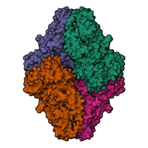
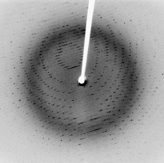
X-ray crystallography remains the primary technique for determining protein and macromolecular complex structures at atomic resolution. By diffracting X-rays through protein crystals and analyzing the resulting diffraction patterns, electron density maps are computed from which complete atomic models are built and refined to Ångström-level precision.
ICDMO's crystallography service covers the complete pipeline — from gene-to-crystal protein production, high-throughput crystallization screening with 2,000+ conditions, and synchrotron data collection at Diamond, ESRF, APS, SSRF, and Spring 8, through structure determination, PDB deposition, and biological interpretation — all under one roof.
Suitable For
Small to medium-sized soluble proteins (MW < 100 kDa preferred)
Proteins forming well-diffracting crystals (enzymes, kinases, receptors)
Membrane proteins via detergent or lipidic cubic phase (LCP) crystallization
Protein–ligand, protein–inhibitor, and protein–cofactor co-crystals
Fragment-based drug discovery (FBDD) soaking campaigns
Protein complexes and multi-subunit assemblies that can be crystallized
Stable proteins without major conformational flexibility in the crystal
Crystallization Process
Three-Phase Crystallization Screening
Our systematic three-phase approach — from initial high-throughput screening to crystal identification — maximizes success even for challenging targets.


Methods
Crystallization Techniques
Vapor diffusion is the core technique in our crystallization platform, implemented via sitting-drop and hanging-drop methods.


Service Workflow
From Gene to PDB Deposition
A systematic, quality-controlled pipeline that has delivered 300+ high-resolution structures across all target classes.
01
01
Crystallization-Grade Protein Production
Gene construct design & codon optimization for target host
Protein expression: E. coli, baculovirus/Sf9, or mammalian HEK-293
Multi-step purification: IMAC affinity, ion exchange, SEC polishing
QC: SDS-PAGE, DLS (polydispersity), SEC-MALS (MW), thermal shift (Tm)
02
02
Initial 96-Well Crystallization Screening
High-throughput sitting-drop and hanging-drop vapor diffusion
2,000+ commercially formulated conditions screened in parallel
Nanoliter dispensing: Mosquito LCP and Crystal Gryphon robots
UV CrysCam fluorescence imaging to identify protein vs. salt crystals
03
03
Optimization of Crystallization Hits
Fine-tuning of pH, precipitant concentration, additive, and temperature
Seeding: streak seeding, micro-seed matrix screening (MMS)
Detergent and lipid additive screens for membrane protein co-crystals
Ligand co-crystallization and soaking with inhibitors or cofactors
04
04
Synchrotron Radiation Data Collection
Remote beamline access: SSRF, Diamond (I03/I04), ESRF (ID23), APS, Spring 8
Cu Kα rotating anode access for preliminary space-group screening
Cryo-loop mounting, flash-cooling in liquid nitrogen, cryo-stream data collection
Multi-wavelength anomalous diffraction (MAD) at tunable beamlines for de novo phasing
05
05
Structure Determination, Modeling & Refinement
Data indexing, integration and scaling with space group determination and data quality statistics
Phasing: Molecular Replacement (Phaser), SAD/MAD (SHELX), MIR (SOLVE)
Atomic model building with iterative restrained refinement against the experimental data
Validation: R/Rfree, Ramachandran and rotamer statistics, clash analysis; deposition support and manuscript figures
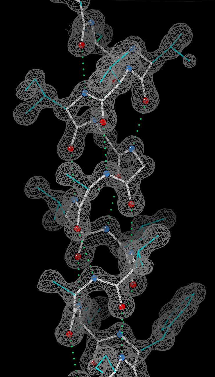
From Crystal to Atomic Structure
The complete X-ray crystallography pipeline — from growing the crystal to depositing refined atomic coordinates in the PDB.
Why Choose ICDMO
Service Advantages
Highest Resolution
X-ray crystallography routinely achieves 1.0–2.5 Å resolution — sufficient to resolve individual atoms, water molecules, and bound ligand geometry with full precision.
Co-Crystal with Ligands
Industry-standard for structure-based drug design: visualize how inhibitors, fragments, and drug candidates bind within the active site at atomic detail.
Synchrotron Access
Established access to major global beamlines (Diamond, ESRF, APS, SSRF, Spring 8) ensures highest-quality diffraction data for even weakly diffracting crystals.
Curated Search-Model Library
A curated library of solved structures supplies search models for Molecular Replacement phasing, cutting time-to-structure for related targets.
PDB-Grade Validation
All structures validated against deposition-standard criteria — Rfree, Ramachandran statistics, rotamer outliers and clash scores — and deposited with full structure factor files for open access.
FBDD Campaign Support
Fragment soaking campaigns against pre-formed crystals: screen 500–1,000 fragments crystallographically to identify novel chemical starting points for lead discovery.
Case Studies
Representative Solved Structures
Services Included
Frequently Asked Questions
Note: Timelines are estimates for well-behaved samples. Challenging targets or low-symmetry crystal forms may require additional optimization cycles. Contact us for a free feasibility assessment before project initiation.
Free Feasibility Assessment
Share your target details and our crystallographers will assess feasibility and recommend the optimal crystallization strategy within 24 hours.
Contact Us Online ConsultationStandard Deliverables
✓Atomic coordinate PDB file
✓Structure factor MTZ file
✓Full geometry and density-fit validation report
✓Detailed refinement statistics
✓Publication-quality figures and viewer sessions
✓Biological interpretation report
✓PDB deposition assistance
✓Manuscript preparation support
Quick Inquiry
Other Structural Techniques
Cryo-Electron MicroscopyNMR SpectroscopySAXS AnalysisStructure-Based Drug DesignStructural Biology OverviewWhat We Need From You
▸Diffraction data, or the protein construct and any crystallisation results to date
▸Sequence, expected oligomeric state and any ligand, cofactor or metal that should be present
▸Search models or homologous structures available, including unpublished internal coordinates
▸The scientific question: binding mode, mechanism, selectivity or conformational state
▸Whether deposition and publication are planned, since that sets the validation standard
How the Work Is Performed
▸Indexing, integration and scaling with space group determination and data quality assessment
▸Phasing by molecular replacement, with search model preparation, or experimental phasing where needed
▸Iterative atomic model building with restrained refinement against the experimental data
▸Ligand fitting with honest assessment of whether the density supports the modelled pose
▸Validation covering R and Rfree, Ramachandran and rotamer statistics, clashes and density fit
▸Structural interpretation directed at the project question, including comparison across related structures
What You Receive
▸Refined coordinates and structure factors at deposition standard
▸Full validation report with the statistics a reviewer or depositor will ask for
▸Ligand density figures showing what the data does and does not support
▸Interpretation tied to your question, with design implications where relevant
▸Publication-quality figures, viewer sessions and deposition assistance
Interpretation and Limitations
Resolution and data quality set a hard ceiling on interpretation. Weak or ambiguous ligand density is reported as such rather than modelled confidently, since an over-interpreted pose can misdirect a chemistry programme for months. Crystal contacts can influence conformation, and a crystallographic state is not always the solution state. Crystallisation and data collection are performed by your laboratory or a partner facility.
Questions We Are Asked Before Scoping
Can you reprocess data that has already been solved?
Yes. Reprocessing and rerefinement frequently improve statistics, correct ligand placement or reveal alternate conformations, particularly for structures solved with older methods.
How do we know the ligand is really there?
By looking at unbiased density and occupancy rather than at the deposited model. We provide the figures that let you judge, and state plainly where the density is ambiguous.
Do you help with deposition?
Yes — validation, metadata preparation and the submission itself, so the structure meets the standards expected by the archive and by reviewers.
What Published Work Reports
The results below were published by independent groups and are cited to show what these computational approaches have achieved in the literature. They are not ICDMO project results, and outcomes vary substantially between targets, datasets and programmes. We do not promise comparable results for any particular project.
Structure prediction now exceeds classical docking on independent benchmarks
On a held-out protein–ligand benchmark, deep-learning complex prediction placed roughly three quarters of ligands within 2 Å of the crystallographic pose, outperforming established docking programs on the same set. For design programmes this changes what can be attempted before a co-crystal structure exists — while leaving binding-mode certainty, and therefore the most consequential chemistry decisions, dependent on experimental structures.
Abramson et al., Nature, 2024 ↗Free energy methods can resolve the differences that decide a synthesis
Published benchmarks on congeneric series report errors near 1 kcal/mol, sufficient to rank analogues that differ by around tenfold in potency. In an integrated design loop this is the calculation reserved for decisions where making the wrong compound costs more than the computation.
Protocol and benchmark studies, Scientific Reports, 2019 ↗