Structural Biology ServicesCryo-Electron Microscopy
Cryo-Electron Microscopy
CryoEM-SPA
CryoEM single particle analysis (SPA) uses the electron scattering mechanism combined with cryogenic freezing, which significantly reduces high-energy electron beam damage to molecular structures, enabling near-native high-resolution structure determination of proteins without crystallization.
✓
Rapid cryogenic sample preparation maintains near-native hydrated state of the sample✓
Enables detection of multiple conformational states of the same target protein✓
No crystallization required — capable of resolving difficult membrane proteins✓
Suitable for determining structures of large protein complexes and assemblies✓
Microgram-level sample requirements with high tolerance for protein purity variation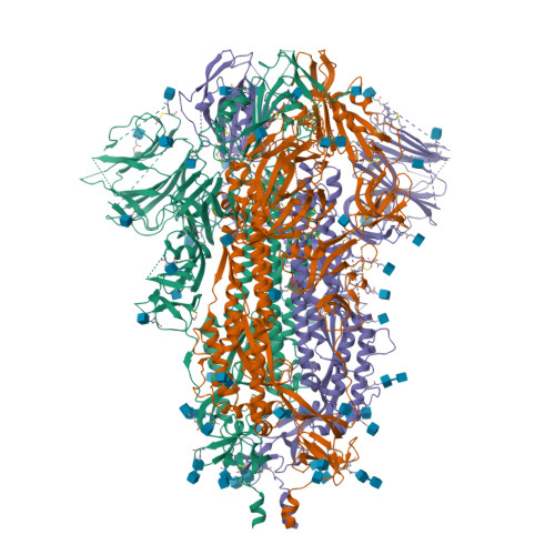
2.5 Å
Best Resolution Achieved
300 kV
Titan Krios Instrument
≤1 Month
Fastest Delivery
0
Crystals Needed
5+
Conformational States / Dataset
Service Advantages
Why Choose ICDMO Cryo-EM Services


Experimental Workflow
From Sample Submission to Atomic Structure
A systematic eight-stage quality-controlled pipeline covering every step from sample receipt through PDB deposition and final report delivery.
How Single-Particle Analysis Works
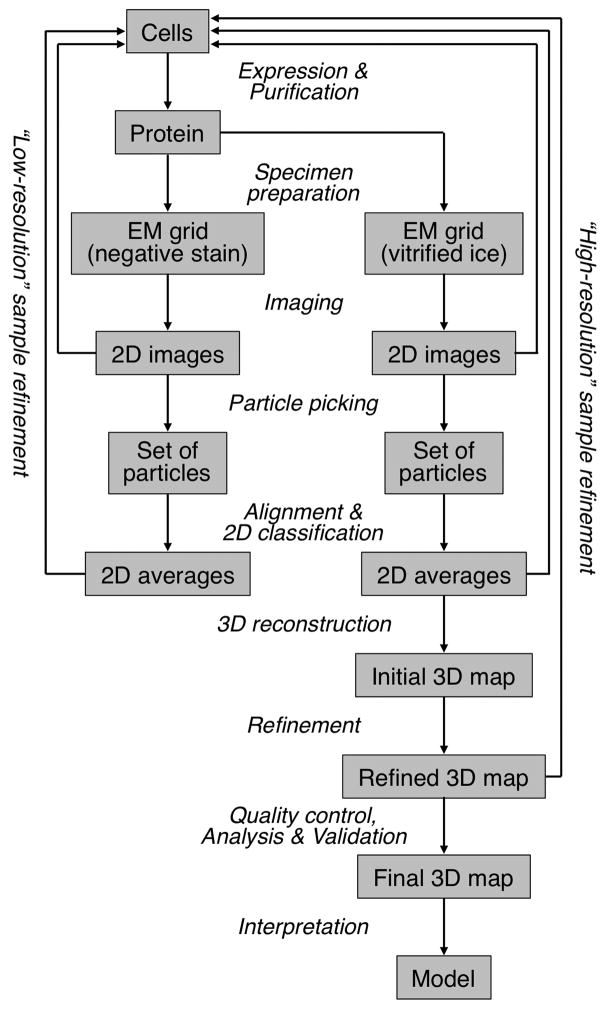
In cryo-EM SPA, tens of thousands of 2D projection images of individual protein particles are collected, computationally aligned, and averaged to reconstruct a high-resolution 3D density map. Unlike crystallography, no ordered crystal is needed — particles are imaged in their near-native hydrated state embedded in vitrified ice.
The process begins with negative-stain TEM for rapid quality assessment, followed by cryo-sample preparation and high-resolution data collection on a 300 kV FEG instrument. Data processing produces density maps with resolution assessed by gold-standard Fourier shell correlation, into which atomic models are then built and refined.
A key advantage is the ability to capture multiple conformational states within a single experiment — 3D classification separates heterogeneous particle populations into distinct structural states, providing a dynamic view of protein function that is inaccessible to crystallography.
01
Sample Preparation
The client provides purified protein samples directly, or specifies the target protein sequence for ICDMO to arrange expression and purification through partner laboratories. Sample purity, concentration, and buffer conditions are assessed and confirmed before proceeding to the next stage.
02
Negative Stain TEM
Heavy metal staining (uranyl acetate or uranyl formate) is applied for rapid quality assessment. Micrographs are analyzed to evaluate particle homogeneity, dispersity, size, shape, and sample concentration — providing a fast go/no-go decision before cryo-sample preparation.
03
Cryo-Sample Preparation
Using a Vitrobot Mark IV plunge-freezer, 3–5 µL of protein sample is applied to a glow-discharged grid. The sample is blotted to form a thin molecular layer and rapidly plunged into liquid ethane (−185°C), vitrifying the sample in amorphous ice to preserve the native protein structure and spatial distribution.
04
Sample Screening & Preliminary Data Collection
Grids are screened using a 200 kV FEG microscope to assess ice thickness, particle distribution, preferred orientation, and 2D class average quality. Preliminary data collection provides resolution estimates and guides optimization of grid conditions before high-resolution acquisition.
05
High-Resolution Data Acquisition
High-resolution data is collected on a 300 kV FEG cryo-electron microscope (Titan Krios) equipped with a direct electron detector (Falcon 4 or K3). Automated acquisition software (EPU, SerialEM) collects tens of thousands to millions of single-particle micrographs with high-throughput automation.
06
Data Processing & 3D Reconstruction
Raw micrograph movies are processed through a multi-step pipeline: beam-induced motion correction, contrast transfer function estimation, automated particle picking, two- and three-dimensional classification, ab initio reconstruction and iterative refinement. Resolution is tracked by gold-standard Fourier shell correlation throughout, and terabyte-scale datasets are handled on dedicated compute.
07
Atomic Model Building
The refined density map is used for atomic model building, with predicted or homologous starting models fitted into density and rebuilt where the map supports it, followed by real-space refinement. Model geometry and fit to density are validated against deposition-standard criteria, and regions not supported by the map are reported as unmodelled rather than filled in.
08
Structure Analysis & Report Delivery
The final atomic structure is analyzed for biological insights: binding interfaces, conformational changes, potential drug-binding pockets, and mechanistic interpretation. A comprehensive project report is delivered including the density map, atomic coordinates, local resolution maps, Fourier shell correlation curves and publication-quality figures.
Representative Case
MRGPRX2 Complex
GPCR ComplexCryo-EM SPASkin Mast Cell
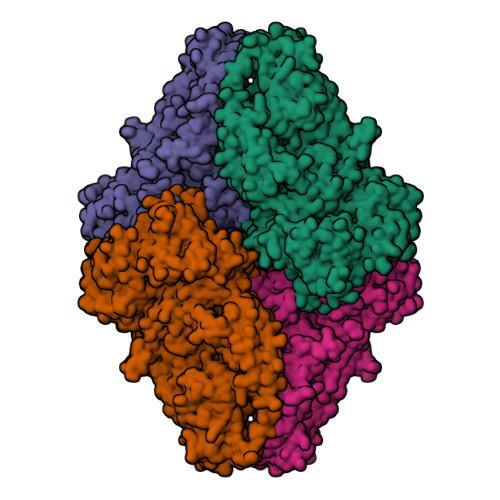
MRGPRX2 – G Protein-Coupled Receptor Complex
MRGPRX2 (Mas-Related G Protein-Coupled Receptor X2) is a GPCR primarily expressed in skin mast cells. It is mainly involved in non-IgE-dependent mast cell activation, playing a key role in Type VII hypersensitivity reactions and pseudo-allergic drug responses.
Using cryo-EM single particle analysis, ICDMO determined the near-atomic resolution structure of the MRGPRX2 complex with its G protein partner. The structure revealed the transmembrane helix packing geometry, key ligand-binding residues in the orthosteric pocket, and a cryptic allosteric site — providing the molecular basis for mast cell activation and a structural template for drug safety profiling and therapeutic development.
Target Class
GPCR (Class A)
Method
CryoEM-SPA
Resolution
3.2 Å
Services Included
Frequently Asked Questions
Note: Timelines are estimates for well-behaved samples. Challenging membrane proteins or heterogeneous complexes may require additional optimization. Contact us for a free negative stain pre-screening before committing to full cryo-EM data collection.
Free Pre-Screening Assessment
Submit your sample for a free negative stain TEM screening. We assess particle quality and provide a resolution feasibility estimate before you commit to full cryo-EM data collection.
Contact Us Online ConsultationStandard Deliverables
✓EM density map in standard map format
✓Fitted atomic coordinate PDB file
✓Local resolution & FSC curves
✓2D/3D class average images
✓Full validation report: geometry, clashes and fit to density
✓Publication-quality figures and viewer sessions
✓Full written interpretation report
✓PDB & EMDB deposition assistance
Quick Inquiry
What We Need From You
▸Sample description: composition, molecular weight, symmetry, expected conformational heterogeneity and purity
▸Raw movies or micrographs where data already exists, with acquisition parameters and detector details
▸Any existing maps, models or homologous structures, including unpublished internal data
▸The scientific question: overall architecture, ligand density, conformational states or interface detail
▸Whether deposition to public databases is planned, since that affects validation and documentation
How the Work Is Performed
▸Motion correction, contrast transfer function estimation and micrograph quality curation
▸Particle picking, two- and three-dimensional classification, and removal of junk and damaged particles
▸Ab initio reconstruction and iterative refinement with resolution tracked by gold-standard correlation
▸Heterogeneity analysis to separate conformational states rather than averaging them together
▸Atomic model building into density, real-space refinement and validation against deposition standards
▸Local resolution estimation and honest annotation of regions the map does not support
What You Receive
▸Density maps in standard format with half-maps and local resolution estimates
▸Fourier shell correlation curves and the full processing record with parameters
▸Fitted atomic coordinates with validation statistics and unmodelled regions identified
▸Class averages and heterogeneity analysis where several states were resolved
▸Publication-quality figures, viewer sessions and deposition support
Interpretation and Limitations
Map quality sets a hard ceiling on interpretation, and no processing recovers information the experiment did not capture. Preferred orientation, air-water interface damage and sample heterogeneity limit achievable resolution and are properties of the sample rather than the software. Regions with weak density are reported as unmodelled instead of built speculatively. Sample preparation, grid screening and data collection are performed by your laboratory or a partner facility.
Questions We Are Asked Before Scoping
Can you process data we already collected?
Yes, and reprocessing frequently improves resolution or resolves conformational states that the original processing averaged together, particularly for older datasets.
What resolution will we get?
It depends on sample behaviour, particle size and symmetry far more than on processing. We give a realistic expectation after seeing initial micrographs rather than a number in advance.
Cryo-EM or crystallography for our target?
Cryo-EM for large, flexible or hard-to-crystallise assemblies; crystallography for smaller, well-behaved proteins where ligand density and higher resolution matter. We advise on your specific target, and sometimes recommend both.
What Published Work Reports
The results below were published by independent groups and are cited to show what these computational approaches have achieved in the literature. They are not ICDMO project results, and outcomes vary substantially between targets, datasets and programmes. We do not promise comparable results for any particular project.
Around three quarters of predicted ligand poses fall within 2 Å
Current deep-learning structure prediction, evaluated on an independent protein–ligand benchmark, placed roughly 76% of ligands within 2 Å of the experimental pose, and reported approximately 80% by coordinate error — a substantial improvement over classical docking baselines on the same set, achieved without supplying a binding site. The corollary matters as much as the headline: roughly a quarter of cases still fall outside that threshold, which is why we report confidence per interface rather than delivering a single model.
Abramson et al., Nature, 2024 ↗