5
Techniques
200+
Structures Solved
1.0 Å
Best Resolution
1–3
Months End-to-End
100%
PDB Deposition
What We Offer
Comprehensive 3D Structure Determination Across Five Platforms
Three-dimensional structure determination lies at the heart of modern drug discovery, mechanistic biology, and protein engineering. Understanding how a protein folds, where substrates and inhibitors bind, and how multi-protein complexes assemble provides the molecular basis for rational therapeutic design.
ICDMO's structural biology platform spans the complete toolkit — from atomic-resolution X-ray crystallography and Cryo-EM through solution-state NMR, rapid SAXS characterization, and fully integrated structure-based drug design. All techniques are delivered under one roof, enabling seamless integrative structural projects.
Suitable For
Drug targets requiring atomic-resolution binding-site characterization
Membrane proteins, GPCRs, and ion channels (Cryo-EM)
Proteins < 50 kDa with dynamic or disordered regions (NMR)
Multi-subunit complexes and assemblies > 200 kDa (Cryo-EM)
Rapid solution-state shape and oligomeric state profiling (SAXS)
Fragment screening and iterative lead optimization campaigns (SBDD)
Mechanism of action studies: substrate, TS, and product complexes
Service Workflow
From Protein to PDB Deposition
01
01
Protein Production
Gene construct design & codon optimization for expression host
E. coli periplasmic, baculovirus/Sf9, or HEK-293 mammalian expression
Affinity (IMAC, Protein A/G) & polishing SEC chromatography
QC panel: SDS-PAGE, DLS, thermal shift assay, SEC-MALS
02
02
Crystallization Screening
High-throughput 96-well sitting-drop & hanging-drop vapor diffusion
2,000+ commercial conditions (Hampton, Molecular Dimensions, Rigaku)
Mosquito and Crystal Gryphon nanoliter dispensing robots
UV CrysCam imaging to distinguish protein vs. salt crystals
03
03
Crystallization Optimization
Fine-tuning pH, precipitant concentration, additives, and temperature
Streak seeding and micro-seed matrix screening (MMS)
Detergent & lipidic cubic phase (LCP) screens for membrane proteins
Co-crystallization and soaking with ligands, inhibitors, or cofactors
04
04
Data Collection & Phasing
Synchrotron radiation beamlines: Diamond, ESRF, APS, SSRF, Spring 8
Cu Kα source access for preliminary space-group screening
Cryo-EM: 300 kV FEG instrument, Falcon 4 / K3 direct electron detector
Phasing by molecular replacement or experimental methods; single-particle reconstruction for Cryo-EM
05
05
Structure Determination & Reporting
Iterative atomic model building with restrained refinement against the experimental data
Stringent validation: Rfree, map correlation, Ramachandran and rotamer statistics, clash analysis
PDB deposition with full structure factor / EM map files
Manuscript-quality structural figures, viewer sessions and a biological interpretation report
Our Techniques
Five Platforms, Full Structural Coverage
Each technique provides unique, complementary information. Our scientists guide you to the right approach for your specific target, timeline, and scientific question.
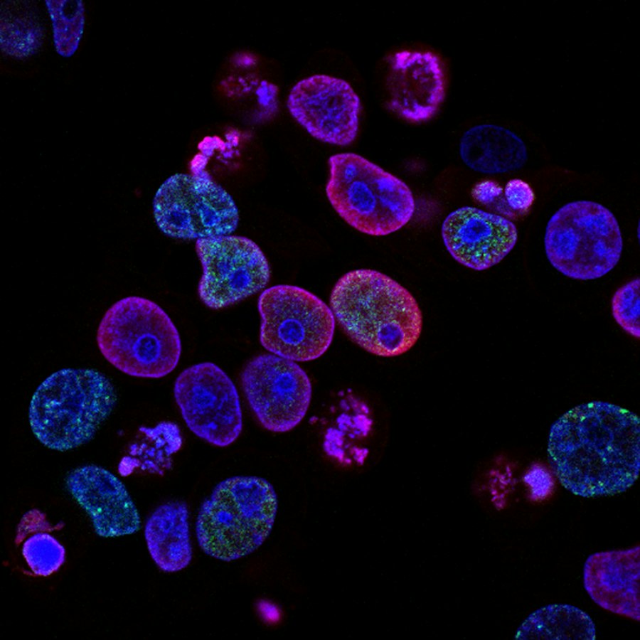
In Detail
Solution-State and Design Services
ICDMO delivers the computational half of structural biology. Data collection is performed by your facility or a partner beamline; we process, interpret and translate the results into structures and design decisions.
NMR Data Interpretation
NMR answers questions crystallography cannot: how a protein moves, which residues a weak fragment touches, and what an intrinsically disordered region is doing in solution. We take acquired spectra and carry out the interpretation — processing, assignment, chemical shift perturbation mapping, relaxation analysis and restraint-based structure calculation — and we state which conclusions the data actually supports.
What we analyse
▸Spectral processing and backbone or side-chain resonance assignment
▸Chemical shift perturbation mapping for binding site and epitope identification
▸Fragment and ligand screening data, including titration series and affinity estimation
▸Relaxation and dynamics analysis across the picosecond to millisecond range
▸NOE and residual dipolar coupling restraints for structure calculation and validation
What you receive
▸Assignment tables and annotated, publication-quality spectra
▸Perturbation maps with the affected residues highlighted on the structure
▸Calculated structural ensembles with restraint statistics and validation
▸Dynamics parameters with the interpretation written out, not only the numbers
▸A statement of which resonances remain unassigned and what that limits
Interpretation quality is bounded by the data: incomplete assignment, spectral overlap in larger proteins and exchange broadening all constrain what can be concluded. We report unresolved regions rather than filling them with assumption. Sample preparation and data acquisition are performed by your laboratory or a partner facility.
SAXS Analysis
Small-angle scattering is the fastest way to learn the solution-state size, shape and oligomeric behaviour of a protein, and the easiest technique to over-interpret. We process scattering curves, assess data quality honestly, derive the structural parameters that are genuinely supported, and fit atomic models or ensembles against the data where the system justifies it.
What we analyse
▸Buffer subtraction, frame assessment, radiation damage and concentration-dependence checks
▸Guinier analysis for radius of gyration, with the fitting range and its validity reported
▸Pair distance distribution, maximum dimension, and Porod and Kratky assessment of flexibility
▸Ab initio shape reconstruction with multiple independent runs and agreement statistics
▸Rigid-body and ensemble fitting of crystal, cryo-EM or predicted models against the curve
What you receive
▸Processed curves with quality assessment and the standard reporting parameters
▸Structural parameters with uncertainty, and an explicit statement of sample monodispersity
▸Shape envelopes or fitted model ensembles with goodness-of-fit statistics
▸Comparison of high-resolution models against the solution data, including where they disagree
▸Figures and a methods section prepared to community reporting standards
SAXS is low resolution and exquisitely sensitive to aggregation and polydispersity. A shape envelope derived from a poor sample is a confident-looking artefact, so data quality assessment comes before interpretation and we will report a dataset as uninterpretable when it is. Measurement is performed by your facility or a partner beamline.
Structure-Based Drug Design
Structure only creates value when it changes what the chemistry team makes next. Our SBDD service runs as a design loop: we analyse the structure and the observed SAR, design and prioritise compounds, apply free energy methods where a synthesis decision warrants the cost, and re-analyse once assay and structural results return. Cycles typically run two to four weeks.
What we analyse
▸Binding mode, interaction geometry, ligand strain and water network in each available structure
▸Growth vectors and unoccupied sub-pockets that analogue design can exploit
▸Selectivity determinants against paralogues and likely off-targets
▸Fragment screening results and how to grow, merge or link confirmed fragments
▸Discrepancies between predicted binding modes and the observed structure-activity relationship
What you receive
▸Prioritised compound designs each cycle with the structural rationale for each
▸Docking, MM/GBSA and FEP results applied selectively according to decision value
▸Updated binding mode models and SAR interpretation as new data arrives
▸Cycle reports comparing what was predicted against what was observed
▸Cumulative documentation suitable for internal review and candidate dossiers
Predictions prioritise hypotheses; they do not establish potency, selectivity or developability. Synthesis and assays are performed by your team or a qualified partner. Where a model repeatedly fails to explain observed SAR, we report that and diagnose the cause rather than retuning until the numbers agree.
Applications
Where Structural Biology Makes the Difference
🔬
Drug Target Validation
Confirm druggability; identify binding pockets, allosteric sites and conformational flexibility for rational drug design.
🧩
Fragment-Based Drug Discovery
Crystallographic screening of 500–1,000 fragment libraries to identify novel chemical starting points for your programme.
📈
Lead Optimization
Iterative co-crystal structures guide SAR: visualize binding modes, optimize key interactions and improve selectivity.
⚙️
Mechanism of Action Studies
Capture substrate, transition-state and product complexes to understand catalytic mechanisms and allosteric regulation.
🔧
Protein Engineering
Rational enzyme variant design, antibody humanization, and protein–protein interface engineering guided by atomic structure.
📋
Regulatory & IP Support
High-quality structures for IND applications, biosimilar comparability studies, and patent binding-mode novelty filings.
Case Studies
Representative Projects


What Published Work Reports
The results below were published by independent groups and are cited to show what these computational approaches have achieved in the literature. They are not ICDMO project results, and outcomes vary substantially between targets, datasets and programmes. We do not promise comparable results for any particular project.
Structure prediction now exceeds classical docking on independent benchmarks
On a held-out protein–ligand benchmark, deep-learning complex prediction placed roughly three quarters of ligands within 2 Å of the crystallographic pose, outperforming established docking programs on the same set. For design programmes this changes what can be attempted before a co-crystal structure exists — while leaving binding-mode certainty, and therefore the most consequential chemistry decisions, dependent on experimental structures.
Abramson et al., Nature, 2024 ↗Free energy methods can resolve the differences that decide a synthesis
Published benchmarks on congeneric series report errors near 1 kcal/mol, sufficient to rank analogues that differ by around tenfold in potency. In an integrated design loop this is the calculation reserved for decisions where making the wrong compound costs more than the computation.
Protocol and benchmark studies, Scientific Reports, 2019 ↗What We Need From You
▸Target sequence and any existing structural data, including unpublished internal structures and maps
▸The scientific question: mechanism, binding mode, conformational state, oligomeric state or design support
▸Available sample information: purity, stability, quantity and any behaviour observed in solution
▸Diffraction images, cryo-EM movies, NMR spectra or scattering curves where data already exists
▸The deadline and the decision the structure has to support, which governs technique selection
How the Work Is Performed
▸Technique selection assessed against target size, flexibility, sample behaviour and the question asked
▸Crystallographic processing, phasing, refinement, ligand fitting and validation
▸Cryo-EM processing: motion correction, CTF estimation, classification, refinement and model building
▸NMR processing, assignment, perturbation mapping and restraint-based calculation
▸SAXS curve processing, parameter extraction, shape reconstruction and model fitting
▸Integrative modelling where several data types must be reconciled into one structural account
What You Receive
▸Refined coordinates with validation reports at deposition standard
▸Maps, processed data and the complete processing record with software versions
▸Structural interpretation directed at your question, not only a set of coordinates
▸Publication-quality figures, viewer sessions and a methods section ready for a manuscript
▸Deposition support for the PDB, EMDB or BMRB where the structure is to be released
Interpretation and Limitations
The resolution and quality of the data set a hard ceiling on what can be concluded, and no processing recovers information the experiment did not capture. Regions with weak density, incomplete assignment or poor scattering are reported as unresolved rather than modelled with false confidence. Sample preparation and data collection are performed by your laboratory or a partner facility; ICDMO delivers the computational and interpretive work.
Questions We Are Asked Before Scoping
Which technique is right for our target?
Crystallography for high resolution where crystals are obtainable, cryo-EM for large or heterogeneous assemblies, NMR for dynamics and small proteins, SAXS for fast solution-state shape. We recommend based on your target and question, and often propose a combination.
Can you process data we already collected?
Yes, and it is common. Reprocessing existing data frequently improves resolution or resolves ambiguity, particularly where the original processing predates current methods.
Can a predicted structure substitute for an experimental one?
For many design decisions, yes. For binding-mode certainty, mechanism claims and publication or regulatory contexts, it cannot. We state which of your intended uses a model can support before you commit to an experimental campaign.
Technique Comparison & Timeline
Note: Timelines are estimates for well-behaved samples. Challenging targets may require additional optimization. Contact us for a free feasibility assessment before project initiation.
Free Feasibility Assessment
Share your target details and our structural biologists will recommend the best technique and provide a no-obligation project plan within 24 hours.
Contact Us Online ConsultationStandard Deliverables
✓Atomic coordinate file (PDB format)
✓Structure factor or EM map file
✓Refinement & validation statistics
✓Publication-quality structural figures and viewer sessions
✓Biological interpretation report
✓PDB deposition assistance